
A New Era in Hip Tendon Repair: Biologic Augmentation with ROTIUM
Greater trochanteric pain syndrome (GTPS) has long challenged surgeons with complex pathology and variable healing outcomes. A new concept for augmentation strategy recently published technique by Dr. Jovan Laskovski in Arthroscopy Techniques demonstrates endoscopic gluteus medius and minimus repair using the ROTIUM® Bioresorbable Wick with a a structural dermal allograft.
Dr. Laskovski, a well-recognized orthopedic surgeon in Akron, OH, has previously utilized ROTIUM in a variety of tendon repair procedures. His work continues to expand the boundaries of biologic healing by adapting proven scaffolding strategies from rotator cuff repairs to hip applications.
Technique Overview
This particular “biologic sandwich” technique integrates two layers of biologic support:
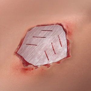
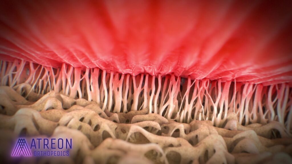
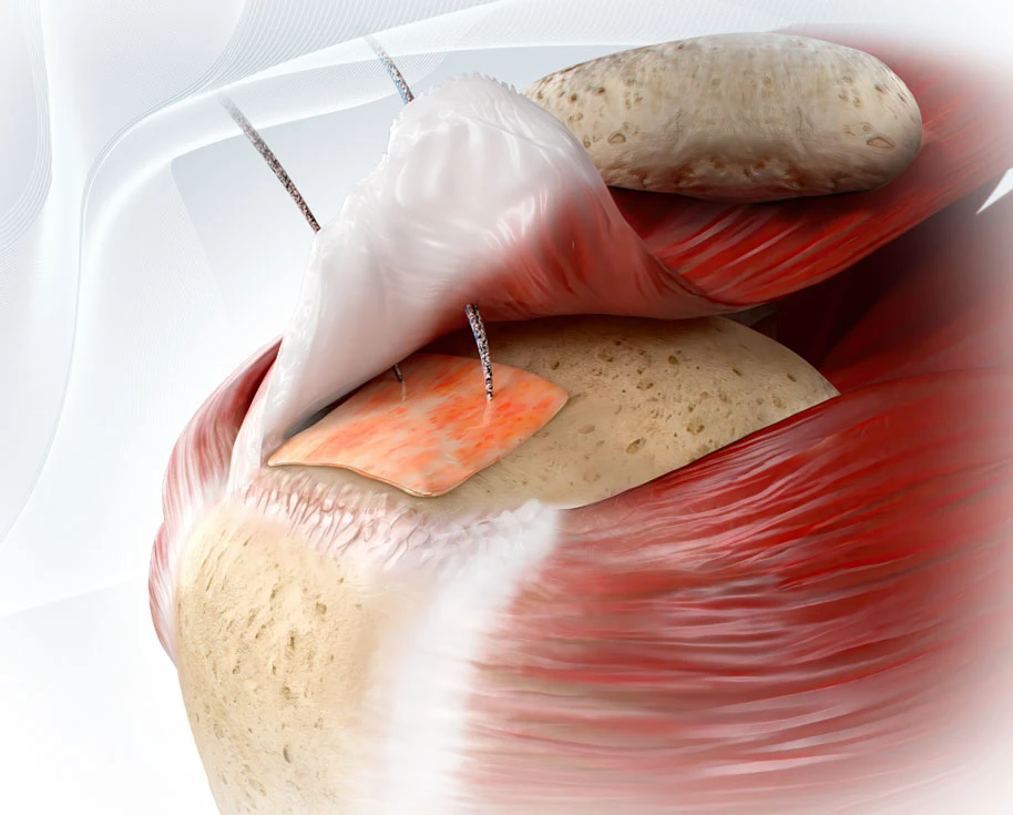
- ROTIUM Bioresorbable Wick: Placed at the tendon-bone interface to mimic the extracellular matrix, wick autologous biology, and support tendon-to-bone healing.
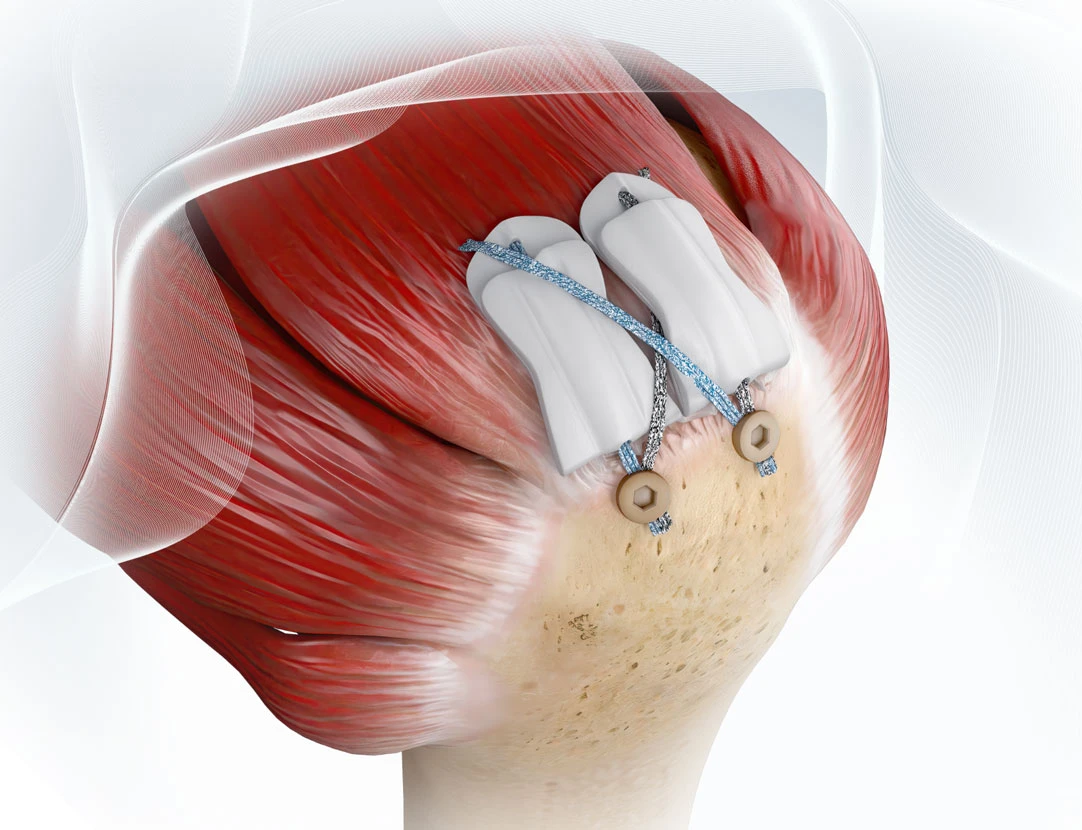
- Acellular Dermal Allograft: Overlaid on the repair to provide structural reinforcement and protect the construct from external stress.
ROTIUM Surgical Technique
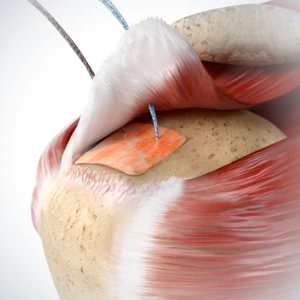
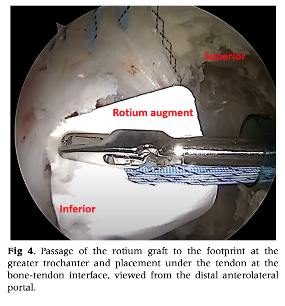
Prepare ROTIUM with a hole and load it over the repair sutures. Shuttle it down the working cannula and position at the tendon-bone interface beneath the gluteus tendons.Retrieve sutures and tie to secure the scaffold in place prior to graft augmentation.
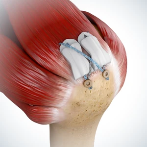
Repair the tendon and compress to the footprint. A structural graft, such as the acellular dermal allograft, may be used as on onlay. This type of “sandwich” technique ensures that the ROTIUM scaffold directly supports biologic healing where it is needed most at the site of tendon-bone integration.
Clinical Rationale
Gluteus medius and minimus tears are biomechanically and biologically similar to rotator cuff tears. The tendon-bone junction in the hip, like in shoulder repairs, frequently presents biological challenges, especially in chronic and revision cases. Studies have shown that scaffolds such as ROTIUM enhance tissue remodeling and improve load-bearing integration at the repair site. This technique adapts those benefits to the weight-bearing demands of the hip.
Efficiency Without Trade-Offs
- Integrates easily with standard hip portals and instruments
- Adds no significant surgical time
- Requires no additional disposables or proprietary tools
Broader Impact: Expanding Indications
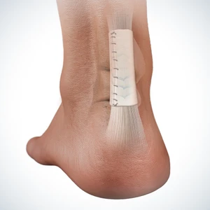
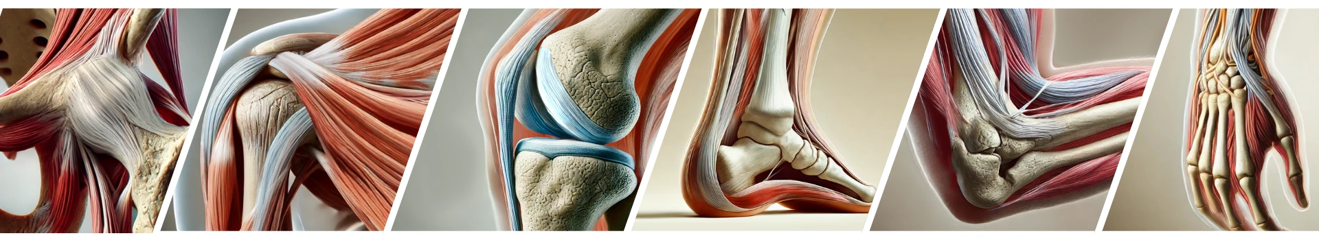
This publication reinforces a major milestone for Atreon Orthopedics: ROTIUM is now cleared for use across a wide range of tendon repairs beyond the shoulder, including hip, knee, elbow, hand, foot, and ankle. With growing adoption, this platform is redefining tendon healing with seamless integration into both open and endoscopic workflows.
Why it matters: Surgeons now have a versatile, biologically active and fully resorbable scaffold to tailor for complex tendon repairs, offering new options for patients with poor tissue quality or high retear risk.