




Patellar Tendon Repair: The Role of Biologic Augmentation Beyond Mechanical Fixation
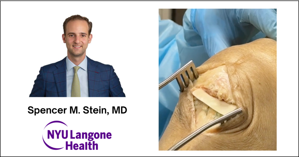
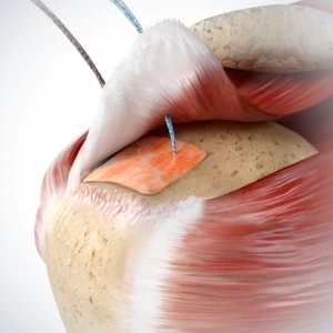
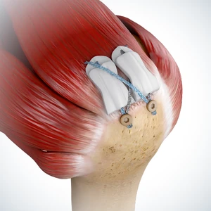
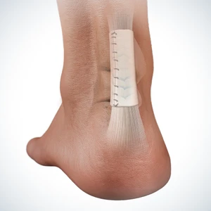
Patellar tendon ruptures are among the most challenging injuries affecting the knee’s extensor mechanism. Although modern fixation techniques provide excellent mechanical stability, surgeons continue to face biologic challenges that can influence tendon healing, rehabilitation, and return to activity (Brinkman, 2024). As the understanding of tendon-to-bone healing evolves, increasing attention is being placed on biologic augmentation strategies designed to support the healing environment—not simply strengthen the repair. In a cadaveric surgical technique, Dr. Spencer M. Stein of NYU Langone illustrates a patellar tendon repair technique utilizing the ROTIUM® Bioresorbable Wick as an interpositional scaffold augmentation placed directly at the tendon-to-bone interface. Understanding Patellar Tendon Injuries The knee’s extensor mechanism includes the quadriceps muscle and tendon, patella, and patellar tendon. Injury to any component disrupts active knee extension, alters gait mechanics, and significantly impacts patient function. Patellar tendon ruptures most commonly affect active patients younger than 40 years and are frequently associated with sports-related injuries. In contrast, quadriceps tendon ruptures are more common in patients over 40 and often result from falls or traumatic events (Taylor, 2024). Several patient factors may increase the risk of extensor mechanism injury, including: Most ruptures occur during an eccentric contraction, when the muscle contracts while lengthening under load. Why Biology Matters After Repair Primary patellar tendon repair generally produces favorable outcomes when performed early. However, returning patients to their previous level of athletic performance remains a challenge. As Dr. Stein explains, delayed reconstruction and revision procedures have consistently less favorable outcomes than primary repair. Gap formation at the repair site and persistent extensor weakness continue to be important clinical concerns. Rather than viewing tendon repair as purely a mechanical problem, surgeons increasingly recognize that successful healing depends on restoring the biologic environment at the tendon-to-bone interface. This concept creates what Dr. Stein describes as a “race between gap formation and healing.” ROTIUM® is designed to support the biologic environment and the body’s natural healing cascade at the tendon-to-bone interface. The Role of Interpositional Biologic Augmentation ROTIUM® Bioresorbable Wick was developed as an interpositional synthetic scaffold designed to support tendon-to-bone healing. Positioned directly between the tendon and its native footprint, the scaffold is intended to support the healing environment by retaining the patient’s own biologic factors at the repair site while providing a bioresorbable scaffold designed to support the body’s natural healing response. The electrospun synthetic scaffold undergoes a biphasic resorption process that releases bioactive degradants, glycolic acid, lactic acid, and caproic acid to support the biologic environment during the normal phases of tendon-to-bone healing. Unlike structural reinforcement patches placed as an onlay, or on top of the tendon, ROTIUM functions as an interpositional scaffold situated directly at the tendon-bone interface, where biologic healing occurs. Featured Surgical Technique: Dr. Spencer Stein, NYU Langone In this cadaveric technique, Dr. Stein performs a primary patellar tendon repair augmented with the 4x3cm ROTIUM® Bioresorbable Wick. Following exposure of the rupture through a standard midline approach, the torn tendon and patellar footprint are carefully prepared before two suture anchors are inserted into the distal pole of the patella. Surgical Technique Overview Step 1: Measure the spacing between the suture anchors and transfer those measurements to the ROTIUM® Bioresorbable Wick. Pass the repair sutures through the scaffold using a free needle. Step 2: Advance the scaffold along the repair sutures until it is positioned at the tendon-to-bone interface adjacent to the patellar tendon. Step 3: Complete the tension-slide repair, reducing the patellar tendon to its anatomic footprint while the ROTIUM scaffold remains interposed at the repair site. Looking Beyond Mechanical Repair As tendon repair techniques continue to advance, biologic augmentation is becoming an increasingly important consideration alongside mechanical fixation. By supporting the healing environment at the tendon-to-bone interface, interpositional scaffold technologies offer surgeons another tool to address one of the most challenging aspects of tendon repair: promoting organized tissue remodeling where durable healing begins. As FDA-cleared indications for ROTIUM® have expanded across any tendon repair applications—including knee and foot & ankle tendon repair—surgeons now have greater flexibility to incorporate biologic augmentation into procedures where optimizing tendon healing is a priority. Conclusion Patellar tendon repair has traditionally focused on restoring mechanical fixation, but today’s surgeons increasingly recognize the importance of the biologic environment at the tendon-to-bone interface. As Dr. Stein’s technique demonstrates, incorporating an interpositional scaffold into a standard repair allows biologic augmentation to be integrated seamlessly into the surgical workflow. As research into tendon healing continues to evolve, biologic augmentation represents another tool surgeons may consider when striving to optimize the environment where tendon-to-bone healing occurs. Learn More Interested in learning more about the ROTIUM® Bioresorbable Wick and its applications in tendon repair? Disclaimer ROTIUM® Bioresorbable Wick is an FDA-cleared bioresorbable scaffold indicated for management and protection of tendons. Individual patient outcomes may vary. Please consult the product Instructions for Use for complete indications, contraindications, warnings, and precautions. References